Equine infectious anemia (EIA) is a potentially life-threatening disease that affects horses worldwide. AffiVET® Rapid Test kits offer a rapid and accurate method for the detection of EIA antibodies in horses. This article highlights the importance of AffiVET® Rapid Test kits in EIA control programs, showcasing their role in preventing the spread of the disease and ensuring equine population health.
First Report of Erysiphe palczewskii Powdery Mildew of Siberian Pea Tree (Caragana arborescens) in Wisconsin and Quebec.
Shoots affected by powdery mildew had been collected from Siberian pea bushes in July 2009 on the University of Wisconsin-Madison campus and on the campus of Université Laval, Quebec City, Quebec. This unique shrub or small tree is occasionally planted in Wisconsin and three shrubs in a bunch that had been affected are the one examples identified on the UW-Madison campus.
In Quebec City, Siberian pea tree is extra generally used as a decorative, usually in hedges (as is the case of the affected vegetation on the Université Laval campus). In each areas, <10% of foliage was visibly affected, however incidence was better on shoots nearer to the bottom than on increased shoots. White-to-grayish mycelium was current on leaves and younger stems and generally utterly lined each higher and decrease leaf surfaces. Dark brown-to-black chasmothecia had been quite a few on leaf blades, petioles, and younger stems, however had been most plentiful on decrease surfaces of leaves.
Morphology of chasmothecia, together with appendages with distinctive terminal dichotomous branching, (1) was in line with descriptions and illustrations of the fungus Erysiphe palczewskii Jacz. (synonym Microsphaera palczewskii) (1-4) considered native to Asia, however often known as an invader of Europe the place it happens on the identical host. For a pattern from Université Laval, imply diameter of chasmothecia was 113 μm, imply appendage size was 185 μm, and barrel-shaped conidia that lacked fibrosin our bodies averaged 30 × 14 μm. Asci contained oval, yellow ascospores with imply dimensions of 20 × 12 μm.
DNA was extracted from chasmothecia, and nuclear rDNA sequences (633 nucleotides) of the Wisconsin (GenBank Accession No. GQ497277) and Quebec (GenBank Accession No. GQ497276) specimens differed by just one nucleotide. The sequences that had been obtained most intently matched GenBank sequences for Oidium spp. (98%) and Erysiphe spp. (97%). Further observations indicated that the identical pathogen affected Siberian pea bushes planted as ornamentals at a number of areas separated by ≥15 km in the metropolitan Quebec space.
This report extends the japanese identified restrict of E. palczewskii in the United States, beforehand identified from collections in Alaska (2), Washington (4), Idaho (4), North Dakota (3), and Minnesota (3). To our information, that is the primary report of this illness in Canada, and it signifies that the distribution of E. palczewskii is transcontinental.
Specimens from Madison, WI and Quebec, QC have been deposited in the U.S. National Fungus Collections (BPI 879152) and the Rene Pomerleau Herbarium of the Canadian Forest Service Laurentian Forestry Centre (QFB-22601). References: (1) U. Braun. Beih. Nova Hedwigia 89:1, 1987. (2) D. A. Glawe and G. A. Laursen. Online publication. doi:10:1094/PHP-2005-1017-01-BR. Plant Health Progress, 2005. (3) D. A. Glawe et al. Online publication. doi:10.1094/PHP-2006-0117-01-BR. Plant Health Progress, 2006. (4) C. Nischwitz and G. Newcombe. Plant Dis. 87:451, 2003.
First Report of Powdery Mildew Caused by Golovinomyces biocellatus on Peppermint in California.
In August of 2009, powdery mildew was noticed on peppermint (Mentha piperita L.) in a number of industrial fields in the Fall River Valley of japanese Shasta County, California. Plant progress was apparently diminished by the illness, however its affect on yield was unknown. White fungal progress was restricted to the adaxial surfaces, the place colonies had been skinny and effused. Heavily contaminated leaves developed a reddish tint as progress prematurely ceased. Doliform conidia ([26.6-] 29.2 [-31.7] × [13.2-] 15.6 [-16.8] μm) had been produced in chains of roughly six conidia.
Foot cells had been cylindrical ([41.3-] 55.2 [-75.0] × [11.2-] 12.0 [-12.8] μm). Immature chasmothecia had been yellowish brown and roughly 100.Zero μm in diameter with flexuous, mycelium-like appendages as much as 200 μm lengthy. All these options had been in line with these of Golovinomyces biocellatus. Asci weren’t noticed. To affirm the identification of the fungus, nuclear rDNA inside transcribed spacer (ITS) areas had been amplified by PCR with common primers ITS4 and ITS5.
The sequence (537 bp) was a precise match for a number of submissions of G. biocellatus in GenBank (e.g., Accession No. EU035602, a sequence of the fungus from mint in Australia [1]). Pathogenicity was confirmed by brushing spores from naturally contaminated leaves onto three rooted cuttings of M. piperita ‘Black Mitchum’. After the vegetation had been lined with a plastic bag for 36 h to keep up excessive humidity, they had been stored on a greenhouse bench at 23 to 28°C.
Three noninoculated vegetation, which served as controls, had been positioned in one other greenhouse in related situations. The experiment was repeated as soon as. All inoculated vegetation developed indicators of powdery mildew inside 7 days of inoculation whereas noninoculated vegetation remained illness free. The fungus on inoculated leaves was morphologically indistinguishable from the one used to inoculate the vegetation. To our information, that is the primary report of G. biocellatus on peppermint in California. References: (1) J. R. Liberato and J. H. Cunnington. Australas, Plant Dis. Notes 2:38, 2007.
Root and Crown Rot of Anthurium Caused by Calonectria ilicicola in Iran.
In the autumn of 2008, a extreme illness of Anthurium andraeanum with wilting and root and crown rot signs was noticed in a greenhouse in the Varamin space of Tehran. A species of Calonectria was remoted constantly from symptomatic tissues on 2% potato dextrose agar (PDA). The fungus produced perithecia and a Cylindrocladium anamorph when incubated on carnation leaf agar beneath near-ultraviolet mild at 25°C. Perithechia had been reddish brown, subglobose to ovoid, and 300 to 400 μm in diameter. Asci had been clavate, hyaline, 90 to 140 × 12 to 19 μm, and tapering to a protracted skinny stalk.

Ascospores had been fusoid, straight to barely curved, 1- (-3) septate, and (30-) 37 to 50 (-65) × (4-) 5 to six.5 (-7) μm (imply = 45 × 6 μm; n = 30). Penicillate conidiophores gave rise to stipe extensions that terminated in sphaeropedunculate vesicles (6-) 7 to 10 (-12) μm in diameter. Conidia had been hyaline, cylindrical, rounded at each ends, straight, (45-) 70 to 82 (-90) × (4-) 5 to six.5(-7) μm (imply = 62 × 6 μm; n = 30), and (1-) 3-septate. On the premise of morphology, the fungus was recognized as Calonectria ilicicola Boedijin & Reitsma.
Koch’s postulates had been fulfilled by spray inoculating 1-month-old seedlings with a conidial and mycelial suspension (105 particles per ml) of the fungus obtained from 14-day-old single-spore colonies grown on PDA at 25°C. Following inoculation, all vegetation had been maintained in plastic baggage in a glasshouse at 25 ± 1°C. After 15 to 25 days, signs resembling these seen in the diseased glasshouse had been detected on inoculated vegetation. C. ilicicola was reisolated from the artificially contaminated tissues.
No signs had been detected on the management vegetation. Nucleotide sequences of the interior transcribed spacer (ITS) areas of the nrDNA operon and the partial histone H3 gene had been decided for derived pressure CPC 16334 as described beforehand (1,3). The ITS sequence (GenBank Accession No. GU057378) matched 100% (644/644 bp) with the sequence of C. ilicicola pressure CBS 463.76 (GenBank AF493963) and the histone H3 sequence (GenBank GU057379) matched 99% (456/458 bp; as a result of two versus three AC repeats in the sequence) with that of C.
[Linking template=”default” type=”products” search=”Anti- Neurofascin (NFASC) Antibody” header=”3″ limit=”122″ start=”4″ showCatalogNumber=”true” showSize=”true” showSupplier=”true” showPrice=”true” showDescription=”true” showAdditionalInformation=”true” showImage=”true” showSchemaMarkup=”true” imageWidth=”” imageHeight=””]
ilicicola pressure CBS 112217 (GenBank AY725686). To our information, that is the primary report of Calonectria and Cylindrocladium genera and the illness brought on by C. ilicicola from Iran. References: (1) R. Cheewangkoon et al. Persoonia 23:55, 2009. (2) P. W. Crous and M. J. Wingfield. Mycotaxon 51:341, 1994. (3) P. W. Crous et al. Stud. Mycol. 50:415, 2004.
Molecular data reveals a new holomorphic marine fungus, Halobyssothecium estuariae, and the asexual morph of Keissleriella phragmiticola
This examine introduces a novel holomorphic marine fungal species, Halobyssothecium estuariae (Lentitheciaceae, Pleosporales), from lifeless Phragmites communis. The new species has semi-immersed, subglobose or ellipsoidal, papillate, conical ascomata, clavate to subcylindrical, quick pedicellate asci and 3-septate, fusoid to ellipsoidal ascospores with rounded ends, pale brown to darkish brown central cells and hyaline finish cells.
The asexual morph has multiseptate, filiform, intercalary, catenate, branched chlamydospores that resemble Xylomyces. The asexual morph of Keissleriella phragmiticola based mostly on mixed LSU, SSU, ITS and TEF1 sequence analyses is reported. The function of molecular identification in delineating cryptic species are additionally mentioned.
First report of Colletotrichum fructicola inflicting anthracnose on Pouteria campechiana in China
Pouteria campechiana (Kunth) Baehni (=Lucuma nervosa A. DC.) is a fruit crop planted in southern China (Gao et al. 2019). It is initially from Central America, and additionally grown there commercially in addition to in some American states (Fadzilah et al. 2018). In March 2019, a leaf spot illness was discovered on P. campechiana in Baoshan, Yunnan, China. Field surveys have been completed in a 0.06 ha orchard in Yunnan Province. Leaf spots have been discovered on 90% of six-year-old vegetation on this subject and have been noticed in different planting areas. The signs initially appeared as small, spherical, brown spots. As the illness developed, the heart of the lesions was sunken with a darkish brown border (Fig. 1).
Under extreme situations, some spots have been joined into bigger irregular spots, and even complete leaves died. The illness severity of completely different vegetation diversified, and some leaves confirmed solely a few brown spots whereas others confirmed many spots. Small fragments of diseased tissues (3×Three mm) have been disinfected in 75% ethanol for 10 s, 1% NaClO for 1 min, and rinsed 3 times in sterilized water.
Then, tissues have been positioned onto potato dextrose agar (PDA), and incubated at 25°C in the darkish for five days. Fungal isolates with comparable morphology have been persistently recovered from diseased tissues. The 25 colonies have been initially cottony, pale white to pale grey on the higher facet and greyish-green with black zonation on the underside of plates.
Conidia have been single-celled and hyaline, aseptate, straight, and cylindrical, with rounded ends (Fig. 1B). The size and width of 200 conidia have been measured for 2 consultant isolates, DHG-1 and DHG-2, and these averaged 14.48 × 5.59 μm and 14.92 × 5.57 μm. Appressoria have been ovoid, generally clavate, brown, averaged 7.47 × 5.86 μm and 7.25 × 5.85 μm (n=30). Brown and globose ascocarp have been noticed on the leaves of Pouteria campechiana.
Asci have been unitunicate, thin-walled, 6-Eight spored, clavate, averaged 51.53×13.01 μm and 50.21 × 13.32 μm (n=30). Ascospores have been hyaline, one-celled, barely curved to curved with obtuse to barely rounded ends, averaged 14.64×5.97 μm and 15.19 × 6.23 μm (n=30). These two isolates have been chosen for molecular identification. DNA was extracted from mycelia with the DNA safe Plant Kit (TIANGEN, Biotech, China).
For additional molecular identification, the inside transcribed spacer (ITS), partial actin (ACT), calmodulin (CAL), chitin synthase (CHS-1), glyceraldehyde-3-phosphate dehydrogenase (GAPDH), beta-tubulin (TUB2), and the Apn2-Mat1-2 intergenic spacer and partial mating kind (Mat1-2) gene (ApMat) genes of the strains (DHG-1, DHG-2) have been amplified utilizing the primer pairs ITS1/ITS4, ACT-512F/ACT-783R, CL1C/CL2C, CHS-79F/CHS-345R, GDF1/GDR1, T1/Bt-2b, and AM-F/AM-R (Weir et al. 2012; Silva et al. 2012), respectively.
The sequences have been obtained and in contrast with GenBank and all of them confirmed over 99% id to the kind pressure of Colletotrichum fructicola ICMP 18581 (Accession nos. JX010165, JX010033, JQ807838, FJ907426, JX010405, JX009866, and FJ917508) (Weir et al. 2012). A phylogenetic tree based mostly on the mixed ITS, ACT, CAL, CHS-1, TUB2, GAPDH and ApMat sequences utilizing the Neighbor-joining algorithm revealed that the isolates have been C. fructicola (Fig. 2). The sequences have been deposited into GenBank with accession MN955541, MN955542, and MN966581 to MN966592.

Pathogenicity assessments have been carried out on eighteen wholesome and tender leaves of six 1-year-old P. campechiana vegetation in a greenhouse. The experiment was repeated twice. The size and width of the inoculated leaves have been between 8-13 cm × 2.5-3.6 cm. The dermis of every examined leaf was evenly scratched in six separate areas with a sterilized needle. Each isolate was inoculated onto at the very least three wounded leaves by inserting 20 μL of a conidial suspension (106 conidia/mL) on the wound websites. Control leaves have been additionally wounded and inoculated with distilled water.
All the vegetation have been then sprayed with distilled water and lined with plastic luggage. After 10 days, preliminary signs appeared as round and deep yellow spots. After a few extra days, the spots grew to become brown, enlarged to as much as 4.Zero mm which was much like signs noticed in the subject, whereas controls remained symptomless. Koch’s postulates have been fulfilled by re-isolation of C. fructicola from diseased leaves, and identification confirmed by sequencing.
Colletotrichum fructicola has been related to anthracnose on mango, apple, pear and cassava (Oliveira et al. 2018). To our data, that is the first report of C. fructicola related to anthracnose of P. campechiana worldwide. These outcomes will present essential info for future epidemiological research and for administration of this illness.
FgPal1 regulates morphogenesis and pathogenesis in Fusarium graminearum
Ascospores are the main inoculum in Fusarium graminearum, a causal agent of wheat head blight. In a earlier examine, FgPAL1 was discovered to be up-regulated in the Fgama1 mutant and vital for ascosporogenesis. However, the organic operate of this well-conserved gene in filamentous ascomycetes isn’t clear. In this examine, we characterised its capabilities in progress, differentiation, and pathogenesis. The Fgpal1 mutant had extreme progress defects and typically displayed irregular hyphal ideas.
It was faulty in infectious progress in rachis tissues and spreading in wheat heads. The Fgpal1 mutant produced conidia with fewer septa and extra nuclei per compartment than the wild kind. In actively rising hyphal ideas, FgPal1-GFP primarily localized to the subapical collar and septa. The FgPal1 and LifeAct partially co-localized at the subapical area in the interdependent method.
[Linking template=”default” type=”products” search=”Tenascin C Antibody” header=”3″ limit=”196″ start=”4″ showCatalogNumber=”true” showSize=”true” showSupplier=”true” showPrice=”true” showDescription=”true” showAdditionalInformation=”true” showImage=”true” showSchemaMarkup=”true” imageWidth=”” imageHeight=””]
The Fgpal1 mutant was regular in meiosis with eight nuclei in creating asci however most asci have been aborted. Taken collectively, our outcomes confirmed that FgPal1 performs a function in sustaining polarized tip progress and coordination between nuclear division and cytokinesis, and additionally it is vital for infectious progress and developments of ascospores by the free cell formation course of. This article is protected by copyright. All rights reserved.
Mycosphaerangium and Neomelanconium (Cenangiaceae) are closest relatives: phylogenetic relationships, morphology and a new species
Based on molecular phylogenetic analyses of a multigene matrix of partial nuSSU-ITS-LSU rDNA, RPB1, RPB2 and TEF1 sequences and by morphological proof, the genus Mycosphaerangium is proven to be the closest relative of Neomelanconium, and confirmed to be a member of the Cenangiaceae (Leotiomycetes). While Mycosphaerangium and Neomelanconium share many traits like comparable conidia, conidiogenesis, asci and ascospores, their apothecia differ notably in excipular options and are subsequently acknowledged as distinct genera.
Mycosphaerangium tiliae, described from North America, is excluded from the genus however proven to characterize the sexual morph of the European Neomelanconium gelatosporum, and it’s subsequently synonymized with the latter. Based on morphology, Neomelanconium deightonii is assumed to be congeneric with Neomelanconium gelatosporum, and it’s lectotypified.
Dermatea tetraspora and Phaeangium magnisporum, the basionyms of Mycosphaerangium tetrasporum and M. magnisporum, respectively, are lectotypified as effectively, and for M. tetrasporum, the asexual morph is recorded for the primary time. Mycosphaerangium quercinum sp. nov. is described as a new species from numerous Quercus hosts in Europe, the place it’s proven to be extensively distributed.
It morphologically and ecologically carefully resembles the North American M. tetrasporum, however differs in paraphysis and ascospore morphology and by croziers at its ascus base. The three accepted species of Mycosphaerangium and the 2 of Neomelanconium are described and illustrated. Mycosphaerangium magnisporum, M. quercinum and M. tetrasporum are recorded to be consistently related to species of Coryneum, indicating a fungicolous behavior, however no proof for fungal associations has been present in Neomelanconium deightonii and N. gelatosporum.
First report of seedling blight of maize brought on by Fusarium asiaticum in Northeast China
Maize [Zea mays L.] is a vital meals and feed crops in northeast of China. In 2019, maize seedling blight with an incidence of as much as 25% was discovered on the subject in Fushun metropolis of Liaoning Province. Typical signs of seedlings have been yellow, skinny, wilt and die. The leaves steadily turned yellow from the bottom of the plant to the highest. Root system was poorly developed.
The major roots have been normally discolored and rotted. And faintly pink or puce-coloured mould was discovered on seeds of the rotted seedings. Symptomatic roots of diseased seedling have been collected and surface-disinfested with 70% ethanol for 1 min and then in 2% NaClO for Three min, rinsed with sterilized water thrice, lower into small items and positioned on potato dextrose agar (PDA) medium for five days at 25 °C. Colonies on PDA have been pink to darkish purple with fluffy aerial mycelium and purple to aubergine pigmentation with the age.
The causal agent was transferred to carnation leaf agar (CLA) medium and incubated at 25°C below a 12-h light-dark cycle. 12 Pure cultures have been obtained from single conidia with an inoculation needle below stereomicroscope. The harvested macroconidia have been hyaline, falcate with single foot cells, 3-5 septate and 28.2- 43.5 μm × 3.7 – 4.9 μm. Chlamydospores have been globose to subglobose (5 to 13.5 μm).
No microconidia have been discovered. The perithecia have been black, ostiolate subglobose. Asci have been hyaline, clavate, measuring 58.1- 83.9 µm × 7.7- 11.9 µm and contained eight ascospores. Morphological characters of the pathogen agreed effectively with descriptions of Fusarium asiaticum (O’Donnell et al.2004; Leslie and Summerell 2006). To verify the identification, partial translation elongation issue 1 alpha (TEF1-a) gene and rDNA inside transcribed spacer (ITS) area of isolate MSBL-Four have been amplified and sequenced (O’Donnell et al. 2015; White et al.1990).
BLASTn evaluation of each TEF sequence (MT330257) and ITS sequence (MT322117), revealed 100% sequence identification with F. asiaticum KT380116 and KX527878, respectively. The isolate MSBL-Four was NIV chemotype as decided by Tri13F/DON, Tri13NIV/R (Chandler et al, 2003) assays. Pathogenicity research have been carried out on maize hybrid “Liaodan 565”. Inoculum of F. asiaticum was ready from the tradition of MSBL-Four incubate in 2% mung beans juice on a shaker (150 rpm) at 25°C for 48 hours.
The 5 liter pots (10 pots) have been crammed with sterilized subject soil and 5 of them have been blended with conidial suspension (300mL in every pot) at 2 × 10⁵ conidia per ml. Ten kernels per pot have been floor disinfected in 2% sodium hypochlorite for five min, rinsed with sterilized water and planted. Five pots have been inoculated and one other uninoculated 5 pots served as controls. The pots have been maintained in a greenhouse at 22-26°C for 40 days. Leaves of the vegetation in inoculated pots have been yellowing and the roots turned discolored or necrotic rot at Four weeks after seedling emergence.

All traits of the illness have been just like these noticed in subject. Non-inoculated management vegetation had no signs. Fusarium asiaticum was reisolated from inoculated vegetation and was an identical to the unique isolate. The experiment was repeated as soon as with comparable outcomes. To our information, that is the primary report of seedling blight brought on by F. asiaticum on maize in northeast China, and it has posed a risk to maize manufacturing of China. References: Leslie J F and Summerell BA. 2006. The Fusarium laboratory guide. Blackwell Publishing, Ames, pp 176-179. O’Donnell et al.2004. Fungal Genetics and Biology 41: 600-623. O’ Donnell et al. 2015. Phytoparasitica 43:583-595. White T J et al. 1990. Academic Press, San Diego, CA, pp 315-322. Chandler E A et al. 2003. Physiological and Molecular Plant Pathology 62(6): 355-367.
Benchmarking an Embedded Adaptive Sampling Configuration Interaction Method for Surface Reactions: H 2 Desorption from and CH 4 Dissociation on Cu(111)
Embedded (emb-) correlated wavefunction (CW) principle allows correct assessments of each ground- and excited-state response mechanisms concerned in heterogeneous catalysis. Embedded multireference second-order perturbation principle (emb-MRPT2) based mostly on reference wavefunctions generated through embedded full energetic house self-consistent subject (emb-CASSCF) principle is at the moment state-of-the-art. However, the factorial scaling of CASSCF limits the dimensions of energetic house and the complexity of programs that may be studied. Here, we assess the efficacy of an alternate CW technique, adaptive sampling configuration interplay (ASCI)-which allows massive energetic areas to be used-for learning floor reactions.
We couple ASCI with density practical embedding principle (DFET) and benchmark its efficiency for 2 reactions: H2 desorption from and CH4 dissociation on the Cu(111) floor. Unlike embedded full energetic house second-order perturbation principle (emb-CASPT2) that precisely reproduces a measured H2 desorption barrier, embedded ASCI, utilizing a very massive energetic house (although one that also contains a small portion of the complete set of orbitals) fails to take action.
Adding an additional correlation time period from embedded Møller-Plesset second-order perturbation principle (emb-MP2) improves the desorption barrier and endothermicity predictions. Thus, the inaccuracy of embedded ASCI comes from the lacking dynamic correlation from the various different electrons and orbitals not included within the energetic house.
For CH4 dissociation, once more embedded ASCI overestimates the dissociation barrier in comparison with emb-CASPT2 predictions. Adding dynamic correlation from emb-MP2 helps appropriate the barrier. However, this composite method suffers from double counting of correlation inside embedded ASCI adopted by emb-MP2 calculations.
[Linking template=”default” type=”products” search=”PE Conjugated” header=”3″ limit=”146″ start=”4″ showCatalogNumber=”true” showSize=”true” showSupplier=”true” showPrice=”true” showDescription=”true” showAdditionalInformation=”true” showImage=”true” showSchemaMarkup=”true” imageWidth=”” imageHeight=””]
We subsequently conclude that the state-of-the-art emb-MRPT2 based mostly on reference wavefunctions generated through emb-CASSCF stays the tactic of alternative for learning floor reactions. emb-ASCI is helpful when massive energetic areas past the restrict of emb-CASSCF are important, resembling to review complicated floor reactions with vital multiconfigurational character (static correlation) however weak dynamic correlation.

Toward a humanized mouse model of Pneumocystis pneumonia
Pneumocystis is a crucial opportunistic fungus that causes pneumonia in kids and immunocompromised people. Recent genomic knowledge present that divergence of main floor glycoproteins could confer speciation and host vary selectivity. On the opposite hand, immune clearance between mice and people is properly correlated. Thus, we hypothesized that humanize mice could present details about human immune responses concerned in controlling Pneumocystis an infection.
CD34-engrafted huNOG-EXL mice managed fungal burdens to a larger extent than nonengrafted mice. Moreover, engrafted mice generated fungal-specific IgM. Fungal management was related to a transcriptional signature that was enriched for genes related to nonopsonic recognition of trophs (CD209) and asci (CLEC7A). These similar genes had been downregulated in CD4-deficient mice in addition to twins with naked lymphocyte syndrome with Pneumocystis pneumonia.
Efficacy of Rezafungin in Prophylactic Mouse Models of Invasive Candidiasis, Aspergillosis, and Pneumocystis Pneumonia
Antifungal prophylaxis is advisable to stop invasive fungal illness attributable to Candida spp., Aspergillus spp., and Pneumocystis jirovecii in sufferers in danger for opportunistic infections, reminiscent of allogeneic blood or marrow transplant recipients, sufferers with hematological illness present process chemotherapy, or sufferers on immunosuppressive therapies.
Current approaches to antifungal prophylaxis require a number of brokers to cowl these key fungi. Rezafungin, a novel echinocandin designed for next-generation properties (e.g., larger stability and long-acting pharmacokinetics for once-weekly dosing), has demonstrated in vitro exercise towards Candida and Aspergillus spp. and efficacy towards Pneumocystis spp. biofilms.
Rezafungin was evaluated in in vivo research of prophylactic efficacy utilizing immunosuppressed mouse fashions of invasive candidiasis, aspergillosis, and Pneumocystis pneumonia. Rezafungin discount of Candida CFU burden was typically larger with growing drug concentrations (5, 10, or 20 mg/kg) and when rezafungin was administered nearer to the time of fungal problem (Days -1,-3, or-5).
Similarly, within the aspergillosis model, survival charges elevated with drug concentrations and when rezafungin was administered nearer to the time of fungal problem. Against P. murina, rezafungin considerably lowered trophic nuclei and asci counts in any respect doses examined.
Rezafungin prevented an infection on the 2 larger doses when put next with automobile and had comparable exercise to the lively management trimethoprim-sulfamethoxazole at human equal doses for prevention. These findings assist Phase Three growth of rezafungin and the potential for single-agent prophylaxis towards invasive fungal illness attributable to Candida spp., Aspergillus spp., and Pneumocystis jirovecii.
The Genus Acervus from Southwestern China and Northern Thailand
Acervus (Pyronemataceae, Pezizales) is a saprobic genus in Pezizomycetes, characterised by coloured apothecia, subcylindrical to cylindrical asci and guttulate ascospores. We collected 4 Acervus samples from China and Thailand. Descriptions and illustrations are launched for all recent samples. One new file of A. globulosus from Thailand, one new species, A. rufus, two recognized species, A. epispartius and A. stipitatus from China are reported.
Phylogenetic evaluation based mostly on 5 genes, the big subunit rRNA (LSU), the interpretation elongation factor-1 alpha (tef1-α), the second largest subunit of RNA polymerase II (rpb2), the biggest subunit of RNA polymerase II (rpb1), and the small subunit rRNA (SSU), revealed the distinct place of the brand new species. The new species is about aside by its crimson apothecia. A key to Acervus species can be given.
A Novel Encochleated Formulation Improves Atovaquone Activity in a Murine Model of Pneumocystis Pneumonia
Although atovaquone is efficient in treating and stopping Pneumocystis pneumonia (PCP), it is use is restricted by nonlinear absorption and hostile occasions. The present examine was undertaken to look at the exercise of encochleated atovaquone (eATQ), a novel lipid-crystal nanoparticle formulation, in a mouse model of PCP. eATQ 100 to 200 mg was superior to commercially accessible atovaquone at 14 days in lowering whole Pneumocystis nuclei and asci. eATQ plus anidulafungin lowered nuclei considerably higher than business atovaquone plus anidulafungin. eATQ is a novel formulation of atovaquone that warrants additional analysis for remedy and prevention of PCP.

Dynamic Sparse Subspace Clustering for Evolving High-Dimensional Data Streams
In an period of ubiquitous large-scale evolving knowledge streams, knowledge stream clustering (DSC) has obtained heaps of consideration as a result of the size of the info streams far exceeds the flexibility of knowledgeable human analysts. It has been noticed that high-dimensional knowledge are normally distributed in a union of low-dimensional subspaces. In this text, we suggest a novel sparse representation-based DSC algorithm, referred to as evolutionary dynamic sparse subspace clustering (EDSSC).
It can address the time-varying nature of subspaces underlying the evolving knowledge streams, reminiscent of subspace emergence, disappearance, and recurrence. The proposed EDSSC consists of two phases: 1) static studying and a couple of) on-line clustering.
During the primary part, a knowledge construction for storing the statistic abstract of knowledge streams, referred to as EDSSC abstract, is proposed which might higher deal with the dilemma between the 2 conflicting targets: 1) saving extra factors for accuracy of subspace clustering (SC) and a couple of) discarding extra factors for the effectivity of DSC.
By additional proposing an algorithm to estimate the subspace quantity, the proposed EDSSC doesn’t have to know the quantity of subspaces. In the second part, a extra appropriate index, referred to as the typical sparsity focus index (ASCI), is proposed, which dramatically promotes the clustering accuracy in comparison with the conventionally utilized SCI index.
[Linking template=”default” type=”products” search=”Anti- ATM Antibody” header=”3″ limit=”196″ start=”4″ showCatalogNumber=”true” showSize=”true” showSupplier=”true” showPrice=”true” showDescription=”true” showAdditionalInformation=”true” showImage=”true” showSchemaMarkup=”true” imageWidth=”” imageHeight=””]
In addition, the subspace evolution detection model based mostly on the Page-Hinkley take a look at is proposed the place the showing, disappearing, and recurring subspaces may be detected and tailored. Extinct experiments on real-world knowledge streams present that the EDSSC outperforms the state-of-the-art on-line SC approaches.
Three Novel Entomopathogenic Fungi From China and Thailand
Entomopathogenic fungi are ubiquitous in tropical rainforests and function a excessive stage of range. This group of fungi not solely has necessary ecological worth but in addition medicinal worth. Nevertheless, they’re typically ignored, and many unknown species have but to be found and described. The current examine goals to contribute to the taxonomical and phylogenetic understanding of the genus Paraisaria by describing three new species collected from Guizhou and Yunnan Provinces in China and Krabi Province in Thailand.
The three novel species named Paraisaria alba, P. arcta, and P. rosea share comparable morphologies as these within the genus Paraisaria, containing solitary, easy, fleshy stroma, fully immersed perithecia and cylindrical asci with thickened caps and filiform ascospores that usually disarticulate at maturity. Phylogenetic analyses of mixed LSU, SSU, TEF1-α, RPB1, RPB2, and ITS sequence knowledge verify their placement within the genus Paraisaria. In this examine, the three entomopathogenic taxa are comprehensively described with colour images and phylogenetic analyses. A synopsis desk and a key to all handled species of Paraisaria are additionally included.
First report of alfalfa leaf spot brought on by Leptosphaerulina australis in China
A illness was noticed on alfalfa cultivar WL168 characterised by white to brown leaf spots of normal to spherical shapes, in Aluhorqin County, Inner Mongolia, China (120°13’23″ to 120°29’14″ E, 43°27’52″to 43°35’16″ N, 281.71m to360.13 m Altitude) throughout 2019 to 2020. The illness primarily offered in spring one month after re-greening and the incidence was 78.30% on this discipline.
Twenty alfalfa vegetation with extreme signs had been used for pathogen isolation. The contaminated tissue was reduce into 2 × 2 mm items, surface-sterilized (in 75% ethanol and 5% business bleach (NaClO) for 30 s and 2 min, respectively), rinsed 5 occasions with sterilized distilled water, and dried between sterile filter paper (Wang et al. 2019). The diseased tissue from every plant pattern had been cultured on potato dextrose agar (PDA) and incubated at 25 °C with 12 h mild/day for ten days.
A fungus was remoted from the diseased leaves at a 100% frequency. Fungal development on PDA was spherical with a black floor, radial edge, and a unclean white middle. The ascocarps had been moved to a clear microscope slide to launch asci and ascospores. Ascocarps had been spheroidal, subglobose brown, 120 to 160 µm × 160 to 180 µm, which include a number of ascus. The dimension of ascus had been 31.zero to 41.6 μm × 75.zero to 87.5 μm and every asci having eight ascospores.
Ascospores had been ellipsoid to rectangular with a gelatinous sheath, brown, 8.Eight to 15.zero µm × 29.9 to 43.zero µm with 2 to three horizontal septums, and zero to 2 vertical septums. A phylogenetic tree was constructed after DNA extraction, PCR with primers to amplify the ITS (VG9: 5′- TTACGTCCCTGCCCTTTGTA-3′ and ITS4: 5′-TCCTCCGCTTATTGATATGC-3′) and LSU (LR7: 5′-TACTACCACCAAGATCT-3′ and LROR: 5′- GTACCCGCTGA ACTTAAGC -3′) areas. The LSU (SUB8273071) and ITS (SUB8218291) amplicons confirmed 99% similarity with L. australis (EU754166.1) within the GenBank.
To confirm the pathogenicity, fungs plugs had been inverted on three compound leaves of 20 alfalfa WL168 for 2 days. Agar plugs (PDA) had been inverted on one other 20 alfalfa WL168 three compound leaves which had been management. All vegetation had been maintained at 22 °C and 44% relative humidity in a development chamber. Similar illness signs had been noticed on contaminated leaves ten days after inoculation, whereas management vegetation confirmed no signs.
The similar fungus was re-isolated from the lesions, and additional morphological characterization and molecular assays, as described above. L. australis has been reported on numerous vegetation, together with Prunusarmeniaca, Dolichos, Poa, Lolium, and Vitis in Australia (Graham and Luttrell., 1961), and additionally from Korean soil in 2018 (Weilan et al., 2018). Additionally, L. briosiana, which is frequent within the USA, China, and different nations, causes Leptosphaerulina leaf spot (Samacet al., 2015). L. trifolii is newly reported to happen in China (Liu et al., 2019). To one of the best of our information, that is the primary report of L. australis infecting alfalfa in China. Considering the big planting space in Inner Mongolia, this pathogen might losses to alfalfa cultivation. Hence, future research ought to discover points of efficient administration of this illness.
Four new species of Talaromyces part Talaromyces found in China
Four new Talaromyces species with none shut kinfolk are reported right here, specifically, T. aureolinus (ex-type AS3.15865 T), T. bannicus (ex-type AS3.15862 T), T. penicillioides (ex-type AS3.15822 T), and T. sparsus (ex-type AS3.16003 T). Morphologically, T. aureolinus is exclusive in producing orange-yellow mycelium and gymnothecia, singly borne asci, and ellipsoidal, spiny ascospores.

Talaromyces bannicus is characterised by the sluggish development price, polymorphic conidiophores, inconsistent stipe lengths, and pyriform to ellipsoidal, echinulate conidia. Talaromyces penicillioides is distinguished by good development and sporulation on malt extract agar (MEA) and yeast extract sucrose agar (YES) media, resembling the colony appearances of sure Penicillium species, and appressed biverticillate and sometimes monoverticillate penicilli bearing globose to ellipsoidal, echinulate conidia.
[Linking template=”default” type=”products” search=”Fscn1 antibody” header=”3″ limit=”116″ start=”4″ showCatalogNumber=”true” showSize=”true” showSupplier=”true” showPrice=”true” showDescription=”true” showAdditionalInformation=”true” showImage=”true” showSchemaMarkup=”true” imageWidth=”” imageHeight=””]
Talaromyces sparsus has extensive, submerged colony margins with sparse aerial mycelium, and conidial areas overlaid with yellow-green, sterile hyphae on MEA medium. These 4 new species are effectively supported by particular person phylogenetic timber based mostly on β-tubulin (BENA), calmodulin (CALM), DNA-dependent RNA polymerase II second largest subunit (RPB2), and inside transcribed spacer area (ITS) gene sequences and the tree of the concatenated BENA-CALM-RPB2 sequence.

miR-495 reduces neuronal cell apoptosis and relieves acute spinal cord injury through inhibiting PRDM5
This research goals to research the position of miR-495 in neuronal cell apoptosis after acute spinal cord injury (ASCI). The ASCI rat mannequin was established and the Basso, Beattie, and Bresnahan (BBB) rating was assessed. miR-495, PR area containing 5 (PRDM5), and Bcl-2 expressions have been measured by qRT-PCR or western blotting. Neuronal cell line PC-12 was subjected to hypoxia situation to simulate the in vitro ASCI mannequin. PC-12 cell apoptosis was measured by movement cytometry, and the interplay between miR-495 and PRDM5 was confirmed by twin luciferase reporter assay.
Results confirmed that BBB rating was considerably decreased in ASCI rats in contrast with sham rats. miR-495 expression was down-regulated in spinal cord tissue of ASCI rats and hypoxia-induced PC-12 cells, and PRDM5 protein stage was up-regulated in spinal cord tissue of ASCI rats and hypoxia-induced PC-12 cells. miR-495 overexpression may cut back apoptosis of PC-12 cells, and up-regulated anti-apoptosis protein Bcl-2 protein stage.
Moreover, PRDM5 was a goal of miR-495, and mRNA and protein ranges of PRDM5 have been negatively regulated by miR-495. miR-495 overexpression may cut back the hypoxia-induced PC-12 cell apoptosis, whereas PRDM5 overexpression abolished this inhibiting impact. The agomir-495 was injected into ASCI rats, and Bcl-2 protein stage and BBB rating have been elevated, however the PRDM5 overexpression reversed these outcomes. Overall, we concluded that miR-495 may inhibit neuronal cell apoptosis and relieve acute spinal cord injury through inhibiting PRDM5.
Second-Order Orbital Optimization with Large Active Spaces Using Adaptive Sampling Configuration Interaction (ASCI) and Its Application to Molecular Geometry Optimization
Recently, chosen configuration interplay (SCI) strategies that allow calculations with a number of tens of lively orbitals have been developed. With the SCI subspace embedded within the imply area, molecular orbitals with an accuracy akin to that of the whole lively area self-consistent area methodology will be obtained. Here, we implement the analytical gradient concept for the single-state adaptive sampling CI (ASCI) SCF methodology to allow molecular geometry optimization.
The ensuing analytical gradient is inherently approximate because of the dependence on the sampled determinants, however its accuracy was adequate for performing geometry optimizations with giant lively areas. To acquire the tight convergence wanted for correct analytical gradients, we mix the augmented Hessian (AH) and Werner-Meyer-Knowles (WMK) second-order orbital optimization strategies with the ASCI-SCF methodology. We take a look at these algorithms for orbital and geometry optimizations, display functions of the geometry optimizations of polyacenes and periacenes, and talk about the geometric dependence of the traits of singlet ASCI wave features.
First report of Erysiphe corylacearum, agent of powdery mildew, on hazelnut ( Corylus avellana) in Romania
Romania has an space devoted to hazelnut (Corylus avellana L.), protecting 890 hectares as of 2019. During October 2020, powdery mildew signs have been noticed on the higher facet of leaves of hazelnut ‘Tonda di Giffoni’ in two business orchards in Dudeștii Vechi, Romania (Fig. 1). The illness was current on 70% of the bushes in planting, with no less than 5 leaves per tree having powdery mildew. Micromorphological examination revealed amphigenous, hyaline, branched, septate mycelial patches of two.
Three to three.6 μm in diameter. Conidiophores measured 24-60 × 5-6 (common: 45 × 6) μm and consisted of erect, cylindrical to flexuous foot cells, adopted by 1-2 shorter cells. Ellipsoid, ovoid to doliform conidia have been produced singly and they measured 19-35 × 16-24 (common: 28 × 19) μm. Chasmothecia have been spherical, 75 to 107 (common: 88) μm in diameter. Nine to 13 straight, generally flexuous, appendages measured 54 to 92 (common: 66) μm in size and that they had 5 occasions dichotomous branched apices with curved suggestions (Fig. 2). Each chasmothecium contained three to 5 ellipsoid, ovoid to subglobose asci measuring 41-58 × 29-55 μm (common 52 × 43) μm.

The asci contained 4 to eight ascospores measuring 13-24 × 11-15 (common 18 × 14) μm. Morphological identification was confirmed by sequencing the ITS-region of rDNA utilizing two isolates from leaves, saved as frozen mycelium at -20°C. PCR was carried out with Erysiphales-specific primer pair PMITS1/PMITS2 (Cunnington et al. 2003). The obtained sequences have been deposited in GenBank (Accession n° MW423075, MW423076).
Blast evaluation of each sequences had 100% identification to ITS rDNA sequences of Erysiphe corylacearum from Azerbaijan (Abasova et al. 2018; Accession n° LC270863), Turkey (Sezer et al. 2017; KY082910), Switzerland (Beenken et al. 2020; MN82272), Iran (Arzanlou et al. 2018; MH047243), Italy (Mezzalama et al. 2020; MW045425) and 99% identification from Georgia (Meparishvili et al. 2019; MK157199).
The sequences had a decrease % identification (83%) to Phyllactinia guttata (Accession n° AB080558) (Fig. 3). Pathogenicity was verified on one-year-old vegetation of C. avellana ‘Tonda di Giffoni’, which have been artificially inoculated with a conidial suspension from contaminated leaves (n = 25). Inoculated vegetation have been incubated at 20 to 28°C with 70 to 80% relative humidity.
White mycelium appeared on the higher floor of the leaves at eight to 10 days after inoculation. No signs have been discovered on management vegetation sprayed with sterile water. The fungus current on inoculated leaves was morphologically an identical to the unique isolates from diseased bushes from the sector. E. corylacearum is native to East Asia and was beforehand reported in Japan on wild species of Corylus (Takamatsu et al. 2015; Accession n° LC009928).
The pathogen more than likely unfold into Europe from east to west of Europe (Heluta et al. 2019), through the Caucasus, ranging from Turkey, Azerbaijan, Georgia, and Iran. P. guttata was thought of the one causal agent of powdery mildew on hazelnut in most international locations, together with Romania (Brown 1995). Compared to P. guttata, which typically develops a mycelium on the underside of leaves, E. corylacearum grows with a white mycelium on the higher facet of the leaves.
[Linking template=”default” type=”products” search=”TNN antibody” header=”3″ limit=”116″ start=”4″ showCatalogNumber=”true” showSize=”true” showSupplier=”true” showPrice=”true” showDescription=”true” showAdditionalInformation=”true” showImage=”true” showSchemaMarkup=”true” imageWidth=”” imageHeight=””]
Recently, E. corylacearum on C. avellana was reported additionally in Ukraine (Heluta et al. 2019), from which it may have moved to Romania. Crop safety methods for hazelnut must be revised in line with the brand new pathogen prevalence.
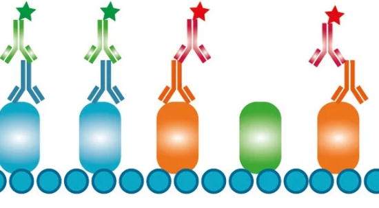
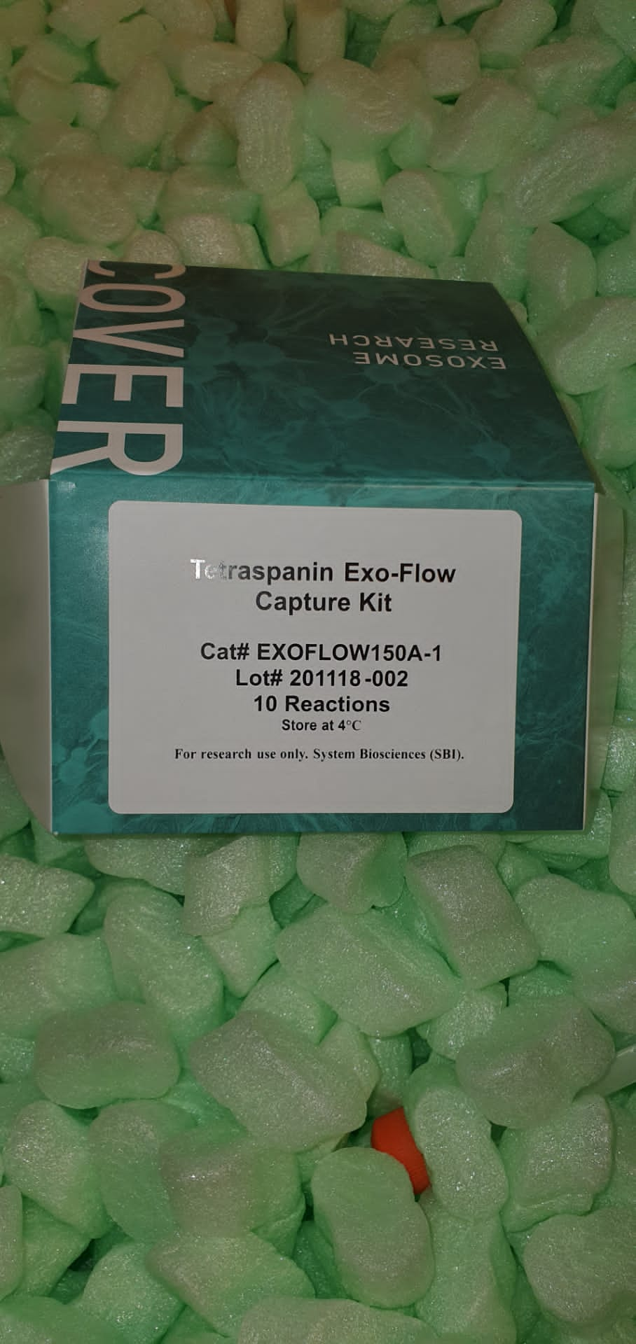
Guide To Selecting Secondary Antibodies
The immunoassays are widely used techniques in both basic research and diagnostic and clinical applications. The suitability of the antibodies used is critical in obtaining the expected result, and this does not only affect the primary antibody (which will bind to the antigen of interest), but also the secondary antibody that will allow us to detect this binding.
Therefore, it is necessary to pay attention to some basic aspects that will help us select the most appropriate secondary antibodies for each case.
Selecting Secondary Antibodies: Criteria To Consider
1. “Host” of the primary antibody:
The secondary antibody must be directed against the species of the primary, so it is essential to know this information. If the primary antibody was produced, for example, in mouse, the secondary one must be an anti-mouse obtained in a different species than this one.
2. Experimental procedure or technique in which it will be used:
This information is essential when choosing the labeling / conjugation of our secondary antibody.
- When detection is carried out by enzymatic reaction, as in the case of techniques such as Western Blot or ELISA immunoassay , secondary antibodies must be labeled with an enzyme (Alkaline phosphatase (AP), Peroxidase (HRP) …) or conjugated to biotin for amplification in two steps.
- When detection is carried out by fluorescence, as occurs in Flow Cytometry (FC), Immunofluorescence (IF), Immunohistochemistry (IHC), Immunocytochemistry (ICC) …, the secondary antibodies must be marked with a fluorochrome.
3. Isotype and subclass of the primary antibody:
The secondary antibody will be directed against the isotype of the primary antibody. Although the vast majority of primary antibodies (especially polyclonal antibodies ) are IgGs, this is an aspect to pay attention to, being essential to know the isotype of the primary antibody, and its subclass is recommended if there is one.
As a reminder, we leave you a summary with the different isotypes of immunoglobulins according to the most frequent species:
4. Purification level of secondary antibody
Secondary antibodies can be presented as an IgG fraction, or as affinity purified antibodies. Each of these two formats has its advantages and disadvantages, which should be known in order to make the most appropriate choice in each case.
- IgG fraction: The main advantage of this presentation is that the high affinity of these antibodies will allow for greater signal amplification, facilitating the reading of the results. In return, the specificity is lower, and may lead to nonspecific junctions that will result in high background noise.
- Affinity purified antibodies: The main advantages in this case would be the high specificity (with the consequent low background level), high sensitivity and high reproducibility of immunoassays. All this to the detriment of affinity, and may lead to weaker reactions.
We hope this short guide has been helpful in helping you select the secondary antibodies that best suit your assay.
Key Factors In The Expression Of Recombinant Proteins
Research in the field of proteomics can cover aspects as diverse as structure, function, modifications, location or interactions between proteins. In any of these fields, a source is usually required from which to obtain a sufficient quantity of the protein of interest for the study.
Chemical synthesis, valid for small peptides , is not a viable option for complete proteins, due to its size and structural complexity. That is why, in the absence of a native protein source, the expression of recombinant proteins is frequently resorted to .
The process of obtaining heterologous proteins basically consists of cloning the gene encoding the protein of interest in an organism other than the one that originally produces it:
However, the expression of recombinant proteins , like any other biotechnological process, is subject to the particularities of each protein in particular, having to solve in each case some challenges such as:
- Solubility
- Conformation
- performance
- Purity
- Additional difficulties for toxic membrane proteins that degrade easily …
Factors That Impact The Expression Of Recombinant Proteins
Countless factors can directly influence viability and performance when expressing proteins recombinantly. Here we summarize some of the most relevant:
1. Sequence : the same protein can be encoded by different sequences forming different codons that give rise to the same amino acid. The variability in the level of expression between two different codons that code for the same amino acid can be up to 250 times. Therefore, before synthesizing the DNA of interest for the expression of our recombinant protein, it is of utmost importance to carry out a codon optimization process that allows us to select the most appropriate sequence.
2. Vector : each of the vector’s structural units can influence the expression performance of a recombinant protein, from the ribosome binding sequences (RBS) to the sequences that encode the tag. But among all of them, the promoter sequence is of special relevance. Note that although generally the stronger the promoter, the greater the performance of the expression, there are exceptions. For example, in the case of toxic proteins, a weak promoter is expected to improve performance.
3. Expression system : the selection of the expression system is key for the expression of the recombinant protein to be successful. This selection should be based on the following criteria:
- Protein characteristics, such as type (membrane, cytoplasmic …), molecular weight, post-translational modifications …
- Application: structural biology, functional assays, antigens for the production of antibodies, protein-protein interaction studies, therapeutic applications …
- Yield to be obtained
- Cost or budget available
We leave you a summary with the strengths and weaknesses of the most common expression systems:
4. Strain (in case of prokaryotes) or cell line (in eukaryotes): once the expression system has been selected, it is necessary to pay attention to the specific strain / cell line to be used, since these can directly influence aspects such as the formation of disulfide bridges or in the reduction of cellular toxicity.
5. Expression conditions : it is also recommended to carry out a small expression test to optimize the different conditions that can affect performance such as the composition of the medium, the temperature, the concentration of the inductors and the induction time or the inoculation volume between others.
The expression of recombinant proteins is a complex process, which requires setting many variables to optimize performance and obtain soluble and functional proteins. We hope that these brief brushstrokes have helped you to clear some doubts.
You can consult our recombinant protein expression service here , and do not hesitate to contact us to ask us any questions in which we can advise you.

Accuracy of Blood Pressure Measurement Devices in Pregnancy: A Systematic Review of Validation Studies.
The correct measurement of blood stress (BP) in being pregnant is important to information medical determination making that impacts each mom and fetus. The intention of this systematic assessment was to find out the accuracy of ambulatory, dwelling, and clinic BP measurement gadgets in pregnant girls.
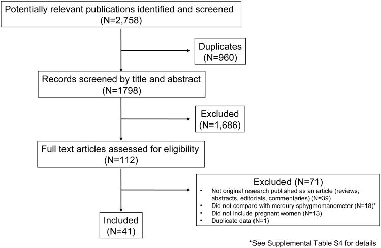
We searched Ovid MEDLINE, The Cochrane Library, EMBASE, CINAHL EBSCO, ClinicalTrials.gov, International Clinical Trials Registry Platform, and dabl from inception by means of August 3, 2017 for articles that assessed the validity of an higher arm BP measurement system in opposition to a mercury sphygmomanometer in pregnant girls.
Two impartial investigators decided eligibility, extracted knowledge, and adjudicated protocol violations. From 1798 potential articles recognized, 41, that assessed 28 gadgets, met the inclusion standards. Most articles (n=32) adopted a normal or modified American National Standards Institute/Association for the Advancement of Medical Instrumentation/International Organization for Standardization, British Hypertension Society, or European Society of Hypertension validation protocol.
Several articles described the outcomes of validation research carried out on>>1 system (n=7) or in>>1 inhabitants of pregnant girls (n=12), comprising 64 pairwise validity assessments. The system was validated in 61% (32 of 52) of research which used a normal or modified protocol.
Only 34% (11 of 32) of the research whereby the system was efficiently validated had been carried out and not using a protocol violation. Given the implications of inaccurate BP measurement in pregnant girls, healthcare suppliers needs to be conscious of and attempt to use the BP measurement gadgets which have been correctly validated in this inhabitants.
Mechanical recanalization in basilar artery occlusion: the ENDOSTROKE research.
OBJECTIVEA research was undertaken to judge scientific and procedural components related to consequence and recanalization in endovascular stroke remedy (EVT) of basilar artery (BA) occlusion.
METHODSENDOSTROKE is an investigator-initiated multicenter registry for sufferers present process EVT. This evaluation contains 148 consecutive sufferers with BA occlusion, with 59% having obtained intravenous thrombolysis previous to EVT.
Recanalization (outlined as Thrombolysis in Cerebral Infarction [TICI] rating 2b-3) and collateral standing (utilizing the American Society of Interventional and Therapeutic Neuroradiology/Society of Interventional Radiology collateral grading system) had been assessed by a blinded core laboratory.
Good (reasonable) consequence was outlined as a modified Rankin Scale rating of Zero to 2 (0-3) assessed after a minimum of Three months (median time to follow-up = 120 days).RESULTSThirty-four p.c had good and 42% had reasonable scientific consequence; mortality was 35%.
TICI 2b-Three recanalization was achieved by 79%. Age, hypertension, National Institutes of Health Stroke Scale scores, collateral standing, and the use of magnetic resonance imaging previous to EVT predicted scientific consequence, the latter Three remaining impartial predictors in multivariate evaluation. Independent predictors of recanalization had been higher collateral standing and the use of a stent retriever.
However, recanalization didn’t considerably predict scientific consequence.CONCLUSIONSBeside preliminary stroke severity, the collateral standing predicts scientific consequence and recanalization in BA occlusion. Our knowledge recommend that the use of a stent retriever is related to excessive recanalization charges, however recanalization by itself doesn’t predict consequence.
The function of different modifiable components, together with the selection of pretreatment imaging modality and time points, warrants additional investigation.